Behavioral Neuroscience, lecture on C start, startle responses in fish
Escape Behavior
IV. Sensory Stimulus for Startle
A. Startle response stimulated by acute vibratory movement of H2O
1. as would happen with a sudden attack
2. can be simulated by a falling golf ball
a. lifting the test tank
b. acoustic stimulation (sound)
3. Mauthner cell inputs from vestibular, auditory & lateral line systems
B. 1o sensory input from vestibular hair cells
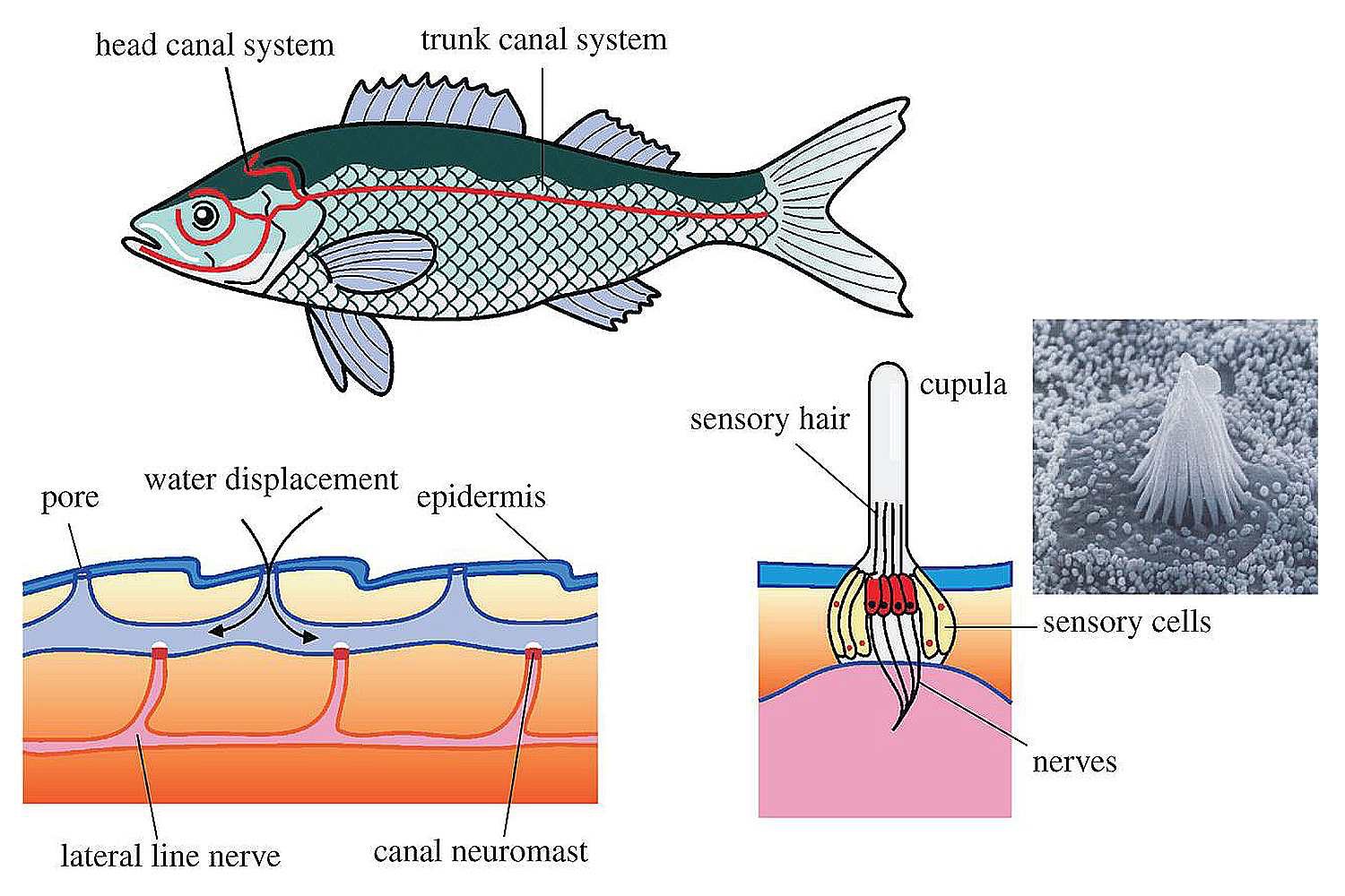 1. Fish hair cells s single Kinocilium
a. at the apical surface
i. does not regress as in mammals
2. with numerous sterocilia
a. embedded in a jelly-like protrusion: the cupula
1. Fish hair cells s single Kinocilium
a. at the apical surface
i. does not regress as in mammals
2. with numerous sterocilia
a. embedded in a jelly-like protrusion: the cupula
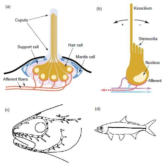 3. hair cell is depolarized when sterocilia move toward the kinocilium
a. hyperpolarized when sterocilia lean away from the kinocilium
4. \ they detect water movements around the body
5. signal travels via VIIIth cranial nerve
C. axons synapse onto Club Endings of Mauthner Cells
1. ipsilateral monosynaptic connection
a. and onto inhibitory interneurons
i. decussates to contralateral Mauthner cell
2. Club Endings: lateral dendrite area of Mauthner Cell
a. mixed Synapse: chemical and electrical
i. rapid, but modifiable transduction of action potential
D. Electrical Synapses
1. Mechanical/electrically conductive link between neurons
a. narrow gap 3.5 nm between presynaptic and postsynaptic cells
i. 20-40 nm for chemical synapse
b. Gap Junctions = channels made from connexins
i. contiguous cytoplasm
ii. ions/current move directly between neurons
iii. voltage of presynaptic cell depolarizes
the postsynaptic cell
(1) no neurotransmitter release/
receptor transduction
(2) \ 10X more rapid - 0.2 ms
(a) chemical synapse 2 ms
E. Chemical synapses = Glu (glutamatergic)
1. Receptors: AMPA/Kainate, NMDA
a. subunits: GluR2/3/GluR5, NR1
2. AMPA-NMDA coupling at synapes via postsyaptic density proteins
a. NMDA current often depends on AMPA depolarization
b. NMDA current augments postsynaptic potentials
c. AMPA up-regulation/trafficking increases NMDA activation
i. makes silent synapses active
F. Mixed synapses - mixed EPSP (excitatory post synaptic potential)
1. rapid depolarization (EPSP) due to 24,000-106,000 gap junction channels
a. central portion of the terminal
b. electrical synapses are bidirectional - do not rectify
i. retrograde spread of dendritic depolarization
(1) to presynaptic club endings
(a) influences excitability
ii. enhances synaptic potential evoked (see c. iii. (1))
c. peripherally surrounding the termimal - chemical release - Glu
i. may be chemically silent
ii. more transmitter release at higher stimulus strengths
(1) electrical cooperativity (see b. ii.)
2. rapid initial current augmented by Glu stimulated EPSP
a. initial depolarization may free NMDA-R of Mg++ blockage
b. AMPA-R and Kainate-R also present
i. also activate NMDA
c. doubly potent chemical component to EPSP
3. Increased stimulus strength increases active terminals
a. 15-20% of total gap channels open usually
b. High frequency stimulation of pVIIIth nerve can evoke LTP
i. includes chemical and electrical component of EPSP
ii. repetitive: 4-6 pulses at 500Hz, every 2 s, for 4 mins
(1) Auditory stimuli in range of 200-800 Hz
4. DA dependent Plasticity of dual synaptic potentials
a. DA release near club ending terminals modulates
1o sensory input to Mauthner cells
b. binds D1 receptors on Mauther cells
i. activates cAMP dependent phosphoylation pathway
ii. phosphoylates Glu receptors, gap junction proteins,
and/or their regulatory molecules
c. Leads to enhancement of mixed EPSP
G. Commissural Inhibitory Interneurons
1. Excitation paired with inhibition of cells on the opposite side
2. Inhibitions mediated by interneurons coupled to the Mauthner axon
3. hair cell is depolarized when sterocilia move toward the kinocilium
a. hyperpolarized when sterocilia lean away from the kinocilium
4. \ they detect water movements around the body
5. signal travels via VIIIth cranial nerve
C. axons synapse onto Club Endings of Mauthner Cells
1. ipsilateral monosynaptic connection
a. and onto inhibitory interneurons
i. decussates to contralateral Mauthner cell
2. Club Endings: lateral dendrite area of Mauthner Cell
a. mixed Synapse: chemical and electrical
i. rapid, but modifiable transduction of action potential
D. Electrical Synapses
1. Mechanical/electrically conductive link between neurons
a. narrow gap 3.5 nm between presynaptic and postsynaptic cells
i. 20-40 nm for chemical synapse
b. Gap Junctions = channels made from connexins
i. contiguous cytoplasm
ii. ions/current move directly between neurons
iii. voltage of presynaptic cell depolarizes
the postsynaptic cell
(1) no neurotransmitter release/
receptor transduction
(2) \ 10X more rapid - 0.2 ms
(a) chemical synapse 2 ms
E. Chemical synapses = Glu (glutamatergic)
1. Receptors: AMPA/Kainate, NMDA
a. subunits: GluR2/3/GluR5, NR1
2. AMPA-NMDA coupling at synapes via postsyaptic density proteins
a. NMDA current often depends on AMPA depolarization
b. NMDA current augments postsynaptic potentials
c. AMPA up-regulation/trafficking increases NMDA activation
i. makes silent synapses active
F. Mixed synapses - mixed EPSP (excitatory post synaptic potential)
1. rapid depolarization (EPSP) due to 24,000-106,000 gap junction channels
a. central portion of the terminal
b. electrical synapses are bidirectional - do not rectify
i. retrograde spread of dendritic depolarization
(1) to presynaptic club endings
(a) influences excitability
ii. enhances synaptic potential evoked (see c. iii. (1))
c. peripherally surrounding the termimal - chemical release - Glu
i. may be chemically silent
ii. more transmitter release at higher stimulus strengths
(1) electrical cooperativity (see b. ii.)
2. rapid initial current augmented by Glu stimulated EPSP
a. initial depolarization may free NMDA-R of Mg++ blockage
b. AMPA-R and Kainate-R also present
i. also activate NMDA
c. doubly potent chemical component to EPSP
3. Increased stimulus strength increases active terminals
a. 15-20% of total gap channels open usually
b. High frequency stimulation of pVIIIth nerve can evoke LTP
i. includes chemical and electrical component of EPSP
ii. repetitive: 4-6 pulses at 500Hz, every 2 s, for 4 mins
(1) Auditory stimuli in range of 200-800 Hz
4. DA dependent Plasticity of dual synaptic potentials
a. DA release near club ending terminals modulates
1o sensory input to Mauthner cells
b. binds D1 receptors on Mauther cells
i. activates cAMP dependent phosphoylation pathway
ii. phosphoylates Glu receptors, gap junction proteins,
and/or their regulatory molecules
c. Leads to enhancement of mixed EPSP
G. Commissural Inhibitory Interneurons
1. Excitation paired with inhibition of cells on the opposite side
2. Inhibitions mediated by interneurons coupled to the Mauthner axon